Location: Heart Model @ 0b039d5e95fd / index.html
- Author:
- Randall Britten <r.britten@auckland.ac.nz>
- Date:
- 2010-09-06 13:29:28+12:00
- Desc:
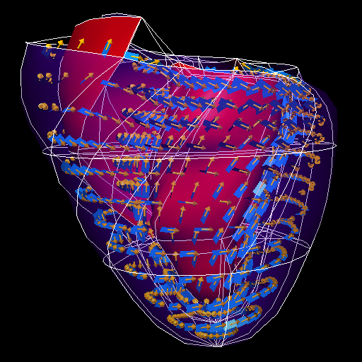
- Demonstration of FieldML 0.2 ? format by Richard Christie, Caton Little and Randall Britten. Representation using FieldML 0.2 ? format of heart model's geometry, converted from of original heart.exnode/heart.exelem dog heart model using a custom conversion which sampled the original 60 element model geometric field. This model consists of 60 elements with RC coordinates interpolated using tri-quadratic Lagrange basis, original used prolate spheriodal coordinates, with 60 bi-cubic-hermite by linear elements. The fibre field has not been converted yet. Files: heart.xml = the fieldml model heart_mesh.connectivity = bulk connectivity data (27 local to global node indexes for each element) heart_mesh.node.coordinates = bulk coordinate parameters (3 RC coordinates per node) fieldml_heart.cmgui = cmgui command file to show (Note: only runs in as yet unreleased development versions of cmgui.) fieldml_heart.png = image of the triquadratic heart
- Permanent Source URI:
- http://models.cellml.org/workspace/heart/rawfile/0b039d5e95fd57f53101623fd2c1c75ea0ff80a8/index.html
We developed a mathematical representation of ventricular geometry and muscle fiber organization using three-dimensional finite elements referred to a prolate spheroid coordinate system. Within elements, fields are approximated using basis functions with associated parameters defined at the element nodes. Four parameters per node are used to describe ventricular geometry. The radial coordinate is interpolated using cubic Hermite basis functions that preserve slope continuity, while the angular coordinates are interpolated linearly. Two further nodal parameters describe the orientation of myocardial fibers. The orientation of fibers within coordinate planes bounded by epicardial and endocardial surfaces is interpolated linearly, with transmural variation given by cubic Hermite basis functions. Left and right ventricular geometry and myocardial fiber orientations were characterized for a canine heart arrested in diastole and fixed at zero transmural pressure. The geometry was represented by a 24-element ensemble with 41 nodes. Nodal parameters fitted using least squares provided a realistic description of ventricular epicardial [root mean square (RMS) error less than 0.9 mm] and endocardial (RMS error less than 2.6 mm) surfaces. Measured fiber fields were also fitted (RMS error less than 17 degrees) with a 60-element, 99-node mesh obtained by subdividing the 24-element mesh. These methods provide a compact and accurate anatomic description of the ventricles suitable for use in finite element stress analysis, simulation of cardiac electrical activation, and other cardiac field modeling problems.
To launch the model, please select 'Zinc Viewer' under navigation on the right.